.jpg)
- Home
- Patients
- Patient Information
- The Spine and MRI Scanning
The Spine and MRI Scanning
What You Need To Know
It is very common that patients will ask for a scan of the spine. It is also very common that a scan is not necessary. A careful history and examination is of far greater value for most simple cases of back pain than a scan.
Magnetic Resonance Imaging Scans (MRI scans) give a very clear picture of the structure of the spine. It does not tell the doctor why the spine is painful. In 85% of patients we are unable to say why back pain occurs. It is often more useful to concentrate on getting back to normal activities and taking good pain killers to get better quicker.
Scans are very helpful in serious spinal disorders such as cancer, infection or nerve compression. There are useful clear guidelines about serious symptoms or ‘Red flags’ that help determine if a scan is required. A previous history of cancer surgery, wieght loss, or weakness in a limb are some of the ‘red flags’.
A common problem for spine surgeons is that of ‘mixed messages’. Patients can sometimes be quite worried by ‘dramatic’ descriptions of what scans show. A loss of water content in a disc makes it look darker than others. This can be normal . It can be described as ‘degenerative discs’ a term that might cause concern, but if it was described as ‘ a bit like getting grey hair’ that would be less frightening. Normal discs will ‘bulge’, and this is not the same as a disc ‘prolapse’. A disc prolapse can sometimes be entirely pain free, but will sometimes cause symptoms. A health care professional who deals with spinal disorders should be comfortable with advising if a scan is required and interpreting your scan in clinical context. They should be able to show you the pictures and explain them to you.
The Detail
MRI (magnetic resonance imaging) is a fairly new technique that has been used since the beginning of the 1980s.
The MRI scan uses magnetic and radio waves, meaning that there is no exposure to X-rays or any other damaging forms of radiation.
There are no known dangers or side effects connected to an MRI scan. The test is not painful; you cannot feel it. Since radiation is not used, the procedure can be repeated without problems. There is a small theoretical risk to the foetus in the first 12 weeks of pregnancy, and therefore scans are not performed on pregnant women during this time.
Because patients have to lie inside a large cylinder while the scans are being made some people get claustrophobic during the test. Patients who are afraid this might happen should talk to the doctor beforehand, who may give them some medication to help them relax.
The machine also makes a banging noise while it is working, which might be unpleasant.
It can give a very clear picture of the structure of the spine.
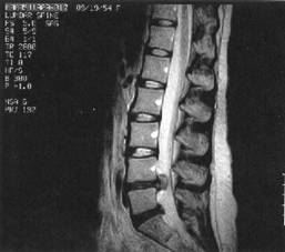
Here you can see a significant disc prolapse at the Lumbar 4/ 5 disc
The patient lies inside a large, cylinder-shaped magnet. Radio waves 10,000 to 30,000 times stronger than the magnetic field of the earth are then sent through the body. This affects the body’s atoms, forcing the electrons into a different position. As they move back into place they send out radio waves of their own. The scanner picks up these signals and a computer turns them into a picture.
The Good News
A scan is very sensitive for the detection of serious spinal disorders such as tumours and infection, such that it will almost always detect this type of disease if it is present. This means it a very useful tool in the assessment and diagnosis of cancer and infection.
Pressure on the spinal cord or on a spinal nerve almost always produces clinical effects that can be found by the person who examines you such as a muscle weakness, altered reflex or areas of changed sensation. A straight leg raise test, for example, if less than 60 degree’s suggests there may be a disc prolapse irritating a nerve.
In older patients there may be spinal stenosis producing narrowing of the spinal canal and difficulty walking. In this situation an MRI scan helps in diagnosis and to plan the treatment options.
If the symptoms are bad enough such that the patient would wish to consider operations or procedures then the scan is the ‘map’ that guides where to direct the treatment.
If the symptoms are not too bad, or improving naturally a scan may not alter the treatment.
The Bits You Might Not Have Heard About
The scan does not tell us where back pain comes from.
In the treatment of back pain rather than nerve pain the scan is used only in patients where surgery is being seriously considered.
Back pain surgery in the form of fusion, disc replacement or other major procedure is only considered after other treatments have been used first. Only a very small number of patients with back pain are suitable for surgery ( some studies suggest less than 3%). Treatments such as exercises and painkillers are safer and often improve symptoms. Recent studies have shown intensive spine exercise and education programs to be as effective as and less of a risk than spine fusion surgery.
Intermittent and variable pains, because they come and go, do not indicate serious spinal disease. Back pain can be very distressing and upsetting for patients. In 85% of cases we are unable to say where the pain is coming from, and a scan doesn’t help.
Spinal specialists are very effective at detecting serious spinal disease and assessing whether a scan is needed.
Surgical scans have a ‘sell by’ date and for surgical procedures need to be current for the condition under treatment. In sciatica surgery for example the scan should be reasonably recent.
MRI scans are very sensitive tests and often show things that are not relevant to current problem. Our philosophy is to ‘treat the man (or woman) and not the scan’.
Simple or 'Non Specific' Low Back Pain
Most National and International guidelines suggest that such conditions do not require an MRI scan.
Most back pain (approx.85%) is non specific, which simply means we do nort know where it comes from. It may be the pain comes from the muscles, joints, discs and ligaments of the spine. In this situation a scan is not helpful and can’t show us the source of the pain. Scans may show changes that are due to natural aging of the spinal column (similar to your hair going grey and thinner as you get older). Almost everyone will have such changes on their scan in middle age and this does not indicate disease. If we incorrectly attribute your symptoms to a normal age related change on the scan we could end up giving you the wrong treatment.
We actually get far more useful information from listening to you, taking a careful clinical history and then examining you. Knowing what you want from seeing us, your expectations, or ‘the agenda’ is very helpful. It helps us to help you.
Why You Might Wait For A Scan
There is a need to ensure the right patients get the right treatment. The priority is for patients with cancer, infection or serious disorders. Patients in this group do not usually wait very long.
Nerve pain and pressure on the spinal cord are conditions that might sometimes require surgery and in a surgical spine clinic also have a priority.
MRI scans are very sensitive and some of the findings we detect we are not certain about the meaning of them. A research study of scans performed in individuals with no back pain at all reported abnormalities in 87% of scans. A common finding is reported as ‘disc degeneration’, it is present in 20% of twenty year olds, 40% of 40 year olds and 80% of 60 year olds. It is a bit like developing grey hair and is nothing to worry about.
It is for these reasons that MRI scans for simple back pain are not usually done and do not have a high clinical priority.
What To Expect If You're Sent For A Scan
A questionnaire is completed concerning the safe use of a scan.
Pacemakers and certain metallic implants can cause serious complications and you should not have a scan
The scan itself is totally painless , but it is quite noisy & ear defenders are given.
If you are claustrophobic there are ‘open’ scanners, or a mild sedative can be given, but have a look at the machine and the room first.
If you can keep very still the scan picture quality is better.